CASE REPORT

A black spot that doesn´t go away after blue laser exposure
PRESENTING AUTHOR
Nancy Arias González
-
Ventura, Marcelo,Bascom Palmer Eye Institute, University of Miami Health System. Pediatric Medical and Surgical Retina. 900 NW 17th St, Miami, FL 33136 EEUU
-
Purpose:
To show an unusual retina finding in a teenage boy. Macular edema is a rare ocular finding after blue laser exposure. The edema resolves but patients may complain of a persistent scotoma. Despite FDA safety standards regulations, it is easy to obtain a class 3B laser online.
-
Case Report:
A 13-year-old male Type 1 diabetic reported to the emergency room complaining of a black spot in the central vision of his right eye (OD). He denied any symptoms in his left eye (OS).
Medical History: Type 1 Diabetes treated with insulin, well controlled.
Upon examination, the patient´s best corrected visual acuity was OD 20/40 and OS 20/20. Slit lamp biomicroscopy findings and intraocular pressures were within normal limits.
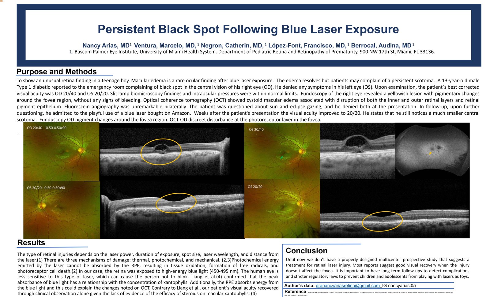
Fundoscopy of the right eye revealed a yellowish lesion with pigmentary changes around the fovea region, without any signs of bleeding. Optical coherence tomography (OCT) showed cystoid macular edema associated with disruption of both the inner and outer retinal layers and retinal pigment epithelium. Fluorescein angiography was unremarkable bilaterally.
The patient was questioned about sun and eclipse gazing, and he denied both at the presentation. In follow up, upon further questioning he admitted to the playful use of a blue laser bought on Amazon. Weeks after the patient's presentation the visual acuity improved to 20/20. He states that he still notices a much smaller central scotoma. Fundoscopy OD pigment changes around the fovea region. OCT OD discreet disturbance at the photoreceptor layer in the fovea. -
Discussion:
The use of high-power lasers has increased in popularity among the general population due to increased accessibility, online availability, and cheaper prices. The unregulated use of this laser has resulted in multiple eye injuries.
The type of retinal injuries depends on the laser power, duration of exposure, spot size, laser wavelength, and distance from the laser.(1) There are three mechanisms of damage: thermal, photochemical, and mechanical. Photochemical energy emitted by the laser cannot be absorbed by the RPE, resulting in tissue oxidation, formation of free radicals, and photoreceptor cell death. In our case, the retina was exposed to high-energy blue light (450-495 nm).(2) The human eye is less sensitive to this type of laser, which can cause the person not to blink. Liang et al.(3) confirmed that the peak absorbance of blue light has a relationship with the concentration of xantophylls. Additionally, the RPE absorbs energy from the blue light, and this could explain the changes noted on OCT.(4,5) Contrary to Liang et al., our patient´s visual acuity recovered through clinical observation alone given the lack of evidence of the efficacy of steroids on macular xantophylls. -
Conclusions:
Until now we don’t have a properly designed multicenter prospective study that suggests a treatment for retinal laser injury. Most reports suggest good visual recovery when the injury doesn’t affect the fovea. It is important to have long-term follow-up to detect complications and stricter regulatory laws to prevent children and adolescents from playing with lasers as toys.
The authors have no financial interests in any material discussed in this article. There are no conflicts of interest to disclose.


