CASE REPORT

Toxocara Chorioretinitis with a Retrolental Membrane: A Case Report
PRESENTING AUTHOR
Sofia E. Parellada, MS
-
Hong-Uyen Hua,University of Miami Miller School of Medicine Bascom Palmer Eye Insititute
-
Purpose:
To describe a case of toxocara chorioretinitis in a 4-year-old boy who presented with a retrolental membrane, masquerading as persistent fetal vasculature (PFV).
-
Case Report:
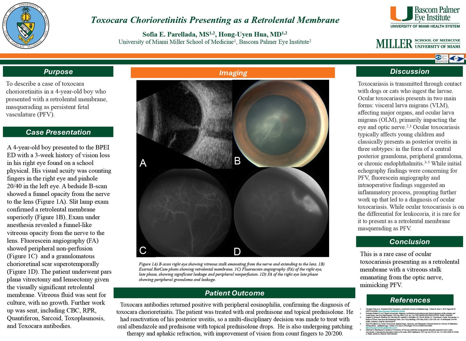
A 4-year-old boy presented with a 3-week history of vision loss in his right eye. His visual acuity was counting fingers in the right eye and pinhole 20/40 in the left eye. A bedside B-scan showed a funnel opacity from the nerve to the lens. Slit lamp exam confirmed a retrolental membrane superiorly. Exam under anesthesia revealed a funnel-like vitreous opacity from the nerve to the lens; fluorescein angiography (FA) showed peripheral non-perfusion and a granulomatous chorioretinal scar inferiorly. The patient underwent pars plana vitrectomy and lensectomy given the visually significant retrolental membrane. Vitreous fluid was sent for culture, with no growth. Further work-up was sent and Toxocara antibodies returned positive with peripheral eosinophilia, confirming the diagnosis of Toxocara Chorioretinitis. The patient was treated with oral prednisone and topical prednisolone, as well as patching therapy and aphakic refraction. Vision has improved from count fingers to 20/200.
-
Discussion:
Ocular toxocariasis typically affects young children and classically presents as a posterior uveitis in the form of a central posterior granuloma, peripheral granuloma, or chronic endophthalmitis. While initial echography findings were concerning for PFV, FA and intraoperative findings suggested an inflammatory process, prompting further work-up that led to a diagnosis of ocular toxocariasis.
-
Conclusions:
This is the first reported case of ocular toxocariasis presenting as a vitreous stalk emanating from the optic nerve disc to a retrolental membrane, mimicking PFV.
The authors have no financial interests in any material discussed in this article. There are no conflicts of interest to disclose.


